Microscopy
Microscopy is a key tool in the study of cell structure. The different types of microscopes, namely light microscopes and electron microscopes, have different applications and allow us to visualize different aspects of cell structure:
Optical Microscopes:
- Use visible light to illuminate specimens. Photons are focused and directed by lenses at the specimen on the stage. Different structures absorb different amounts and wavelengths of light. The photons pass through the thin specimen and enter the eye through more lenses.
- Can magnify up to 1500 times.
- Allow us to see the overall shape and structure of cells, as well as some organelles such as the nucleus and mitochondria.
- Produce coloured photomicrographs, and differential staining can be used to distinguish between cells or organelles.
- Live specimens can be used.
- Maximum resolution is 0.2 $\mu m$.
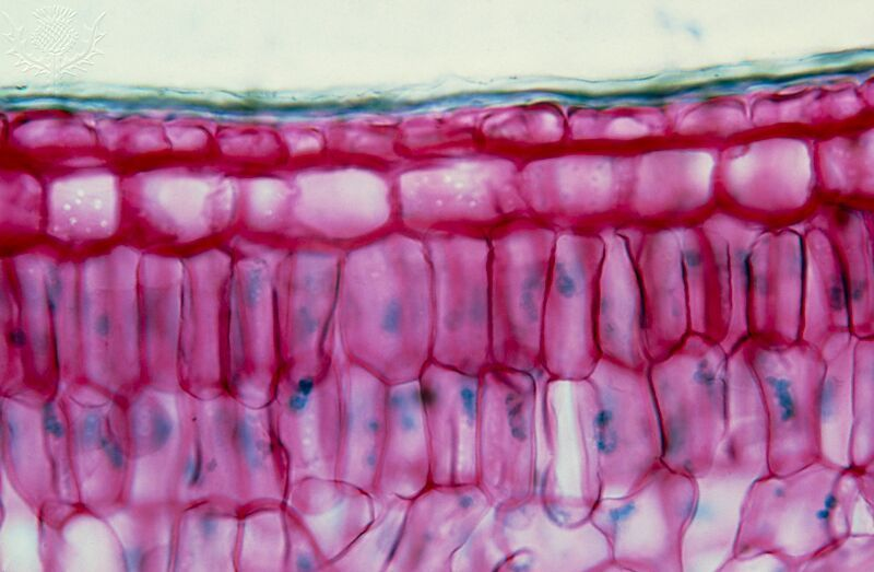
Photomicrograph of a transverse section through a holly leaf (Encyclopædia Britannica)
Electron Microscopes:
- Use beams of electrons to illuminate specimens.
- Can magnify up to 50 000 000 times.
- Allow us to see much greater detail of cell structure, including individual organelles and macromolecules such as proteins and nucleic acids.
- Produce black and white electron micrographs due to the lack of photons. Electron micrographs can be artificially coloured post-capture. This produces a false-coloured electron micrograph.
- Specimens must be dead, since they sit in a vacuum. This is to cause as little unnecessary obstruction to the beam of electrons as possible.
- Maximum resolution is 0.2 $nm$.
There are two main types of electron microscopes. These are Transmission Electron Microscopes (TEM) and Scanning Electron Microscopes (SEM). It is important to know the difference between these two, as they have different applications:
Transmission Electron Microscope:
- Use electromagnetic lenses to focus a high energy beam of electrons through a thin specimen. Different structures absorb different amounts of the beam. Denser structures absorb more electrons. The resultant beam is focused onto a fluorescent screen, producing a black and white micrograph with denser structures appearing darker.
- Allow us to see the internal ultrastructure of cells and their organelles in great detail.
- Can be used to study the structure of viruses and macromolecules such as proteins and nucleic acids.
- Produces a two-dimensional micrograph.
- Can magnify up to 2 000 000 times.
- Maximum resolution of 0.5 $nm$.
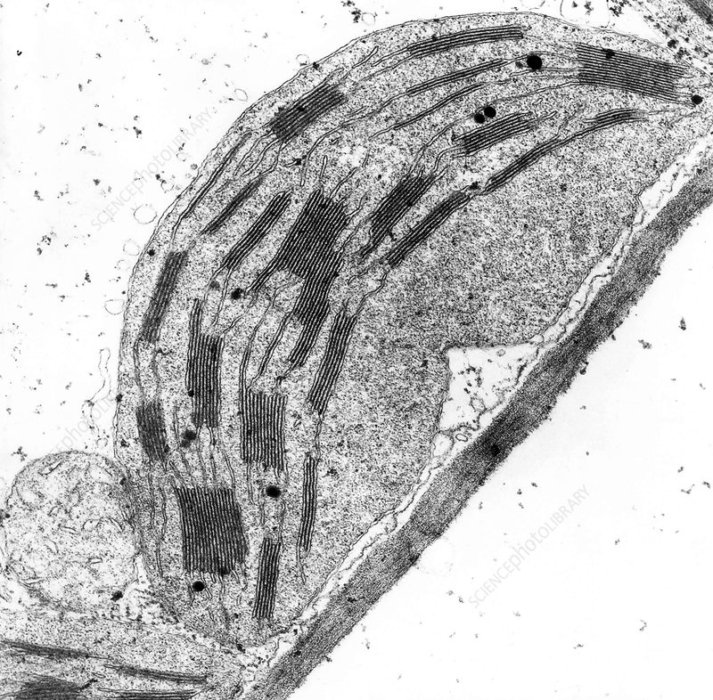
TEM micrograph of a chloroplast (Science Photo Library, Christiane Tuquet)
Scanning Electron Microscope:
- Use electromagnetic lenses to focus a high low energy beam of electrons onto the surface of the specimen. Reflected electrons are detected by a collecting device and amplified to produce a black and white micrograph.
- Produce three-dimensional micrographs of cell surfaces and surface structures of organelles.
- Can be used to study cell surface morphology and the structure of tissues and organs.
- Can magnify up to 500 000 times.
- Maximum resolution of 3-10 $nm$.
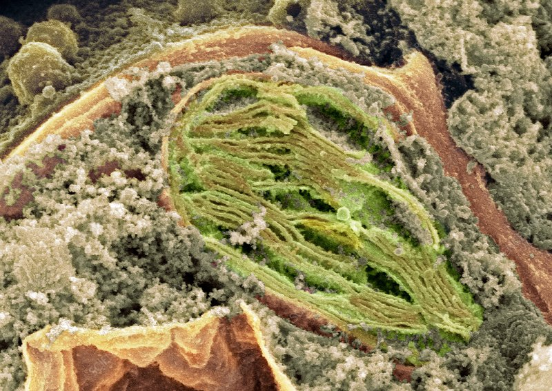
SEM micrograph of a chloroplast (Science Photo Library, David Furness)
Laser Scanning (Confocal) Microscope
One final type of microscope is the confocal microscope, which uses lasers to scan the surface of a specimen. The lasers are focused using objective lenses to a single point on the specimen’s surface. This beam is then reflected back into a photomultipliear tube, which amplifies the signal onto a detector. These pixels are arranged in order by a computer, and the image is displayed on a computer screen.
Confocal microscopes have a high resolution and contrast, and can show coloured micrographs. They also have depth selectivity, meaning a specific structure within a specimen can be targeted.
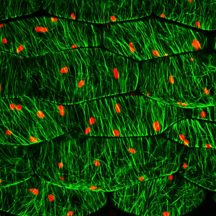
Confocal micrograph of a leaf. Microtubules shown in green and Chloroplasts shown in red. (Unknown Source)
Magnification and Resolution
Magnification is defined as:
The number of times larger an image appears, compared with the actual size of the object.
This means that, due to the linear magnification achieved by microscopes, if a specimen is magnified 100 times, it appears to be 100 times wider and 100 times longer than it really is.
Magnification can be calculated using the equation $I = AM$; where $I$ is the image size, $A$ is the actual size of the specimen, and $M$ is the magnification.
Note: when using this equation, image size and actual size must be in the same units.
Resolution is defined as:
The clarity of an image; the ability to distinguish between two distinct points.
This means if two distinct points cannot be resolved, they are closer together than the resolving power (resolution) of the instrument, and appear as a single point.
For example, a ribosome is about 20 $nm$ in diameter, whereas a light microscope’s resolution is only 200 $nm$, so a ribosome is not visible on a light micrograph.